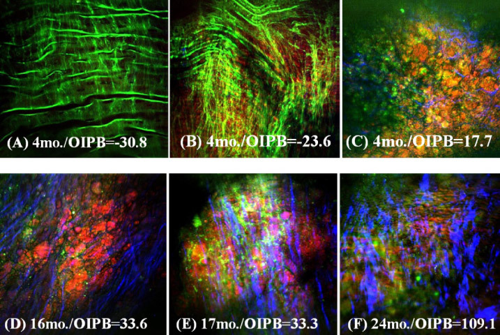
Fig. 10.

Representative epi-NLO images obtained from the luminal surface of thoracic aorta of rabbits sacrificed at (A), (B) and (C) 4 months (D) 16 months (E) 17 months and (F) 24 months of age. Images were collected using 20x air objective lens. These are the sum images of 5 consecutive image planes taken at 2 µm steps from 20µm to 30µm beneath the luminal surface. Green is TPEF signal representing elastin fiber and other endogenous fluorescent molecules; Blue is SHG signal representing fibrillar collagen; Red is CARS signal representing lipids-rich structures in plaque.
