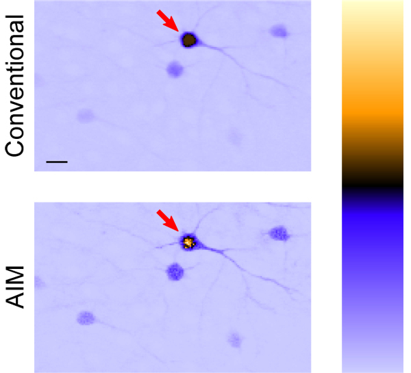
Fig. 6.

Image of GFP-labeled mouse neurons; conventional TPEF (top) cannot properly quantify what should be a bright neuron body indicated by arrow due to saturation. Saturation is avoided and the fluorescence of the neuron is properly captured in the linearized AIM image (bottom) without sacrificing SNR for dim objects. Arbitrary units of fluorescence are represented by a color look-up table (right) to highlight this effect; the bottom of the bar is zero fluorescence. Scale bar is 20 μm.
