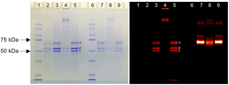
Fig. 2.

SDS-PAGE gel images showing Coomassie blue staining (left panel) and NIR fluorescence (right panel) for the same gel. Lanes 1 and 6 are protein MW standards. Lane 2 is unlabeled hFg only. Three major bands (from top to bottom) represent α (63.5 kDa), β (56 kDa), and γ (47 kDa) chains, respectively. Lanes 3–5 and 7–9 are protein with NIR labels (3–5: hFG and 7–9: BSA) Lanes 3 and 7 have no TG2. Lanes 4 and 8 are incubated with TG2. Lanes 5 and 9 are incubated with both TG2 and EDTA. EDTA inhibits the cross-linking reaction of TG2.
