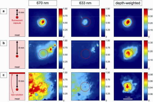
Fig. 2.
Color-coded NIRF images of the tissue phantom study. Capsules containing NIRF dye Cy5.5 embedded in meat as sketched in a vertical view in the first column. Images with illumination at 670 (left) or 633 nm (middle), and the resulting depth-weighted images (right) are shown. The red circles indicate the position of the fluorescent target. (a) capsule located 4 mm below the surface. (b) capsule located 8 mm below the surface. The asterisk indicates an area of strong superficial tissue autofluorescence, which is strongly reduced in the depth-weighted image. (c) capsule located 8 mm below the surface and a dye solution is applied topically on the surface. The color bars represent relative fluorescence intensities (arbitrary units).

