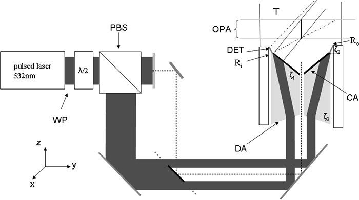
Fig. 2.

schematic experimental setup for dual mode scanning acoustic microscopy (DSAM): ring shaped illumination (IA) for the pulse echo-mode, free beam illumination (IP) for the photoacoustic mode; T: water filled tank; DET: piezoelectric ultrasound detector; CA: black coated axicon; DA: divergent optical axicon; OPA: operating area of the DSAM; Ri and Ro: inner and outer radii of the ultrasound detector.
