Figure 3.
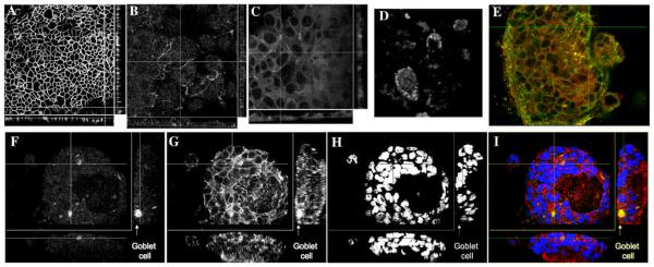
Ptk6 null cells polarize and differentiate on filters and form organoids in collagen.
PTK6 cells were plated on Transwell filters (A-C) or were plated in collagen as a single cell suspension (E-I) and were allowed to grow into organoids. Shown are confocal horizontal (XY; D-I) and vertical sections (XZ and YZ; A-C, F-I) through the filters and an organoid. A, Cells on filters stained for E-cadherin, 40X objective 2X zoom. B, Cells on filters stained for occludin, 40X objective. C, Cells on filters stained for ZO1, 40X objective. D, Cells in an organoid stained for chromogranin, 60X objective 4X zoom. E, Organoid stained for villin (red), and β-catenin (green), 60X objective 1.5X zoom. F, Organoid stained for mucin2,), G, β-catenin (red), H, DAPI, blue) or I, overlay of all three 60X objective.

