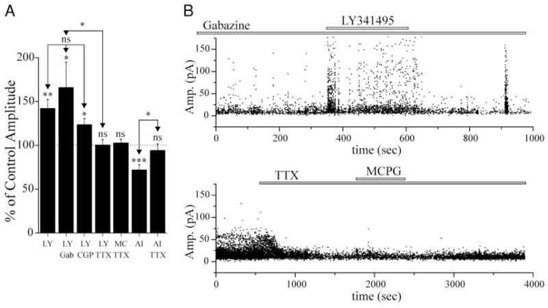
FIG. 4.
Changes in sEPSC amplitude elicited by mGluR antagonists require action potential generation, but not involvement of GABAergic cells. A: graph showing the effect of LY341495 (2 μM), MCPG (100–200 μM), or AIDA (1 mM) on sEPSC amplitude under several conditions. The enhancement of sEPSC amplitude by LY341495 was not affected by concomitant exposure of slices to antagonists for either GABAA (gabazine, 8 μM, n = 8), or GABAB receptors (CGP55845, 0.2–1 μM, n = 8). LY341495, MCPG, or AIDA perfusion had no significant effect on sEPSCs in the presence of 1–2 μM TTX (LY341495: n = 6; MCPG: n = 7; AIDA: n = 6). *, a significant increase in sEPSC amplitude compared with control (P < 0.05). The effects of LY341495 alone on sEPSC amplitude were not significantly different from those obtained in gabazine or CGP55845. LY, LY341495; MC, MCPG; AI, AIDA; Gab, gabazine; CGP, CGP55845. B: graphs of sEPSC amplitude vs. time for 2 cells included in the bar graph of A. Top: cell was exposed to gabazine (8 μM) for >15 min before application of LY341495 (2 μM). Note, the burst of EPSCs at ~900 s, is likely due to a spontaneous epileptiform event. Bottom: cell exposed to TTX (2 μM), followed by MCPG (0.2 mM) ~1,500s later. ●, a single sEPSC.

