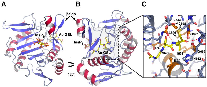
Figure 3. Structure of activated TcdB CPD bound to Ac-GSL-AOMK inhibitor.
(A) Ribbon structure of TcdB CPD in complex with InsP6 viewed from above the InsP6 binding site. InsP6 is shown as a stick model.
(B) A view of the structure rotated ~120º to show inhibitor bound in the active site. InsP6 and the inhibitor are shown as stick models. The β-flap hairpin that separates the InsP6 binding and active site is indicated.
(C) Close-up view of the substrate binding pocket. Hydrophobic residues in the S1 binding pocket are shown as orange sticks, and the inhibitor is shown as yellow sticks. Sidechains that interact with the P1 leucine are shown; hydrogen bonds are indicated by dotted lines.
See also Figure S2.

