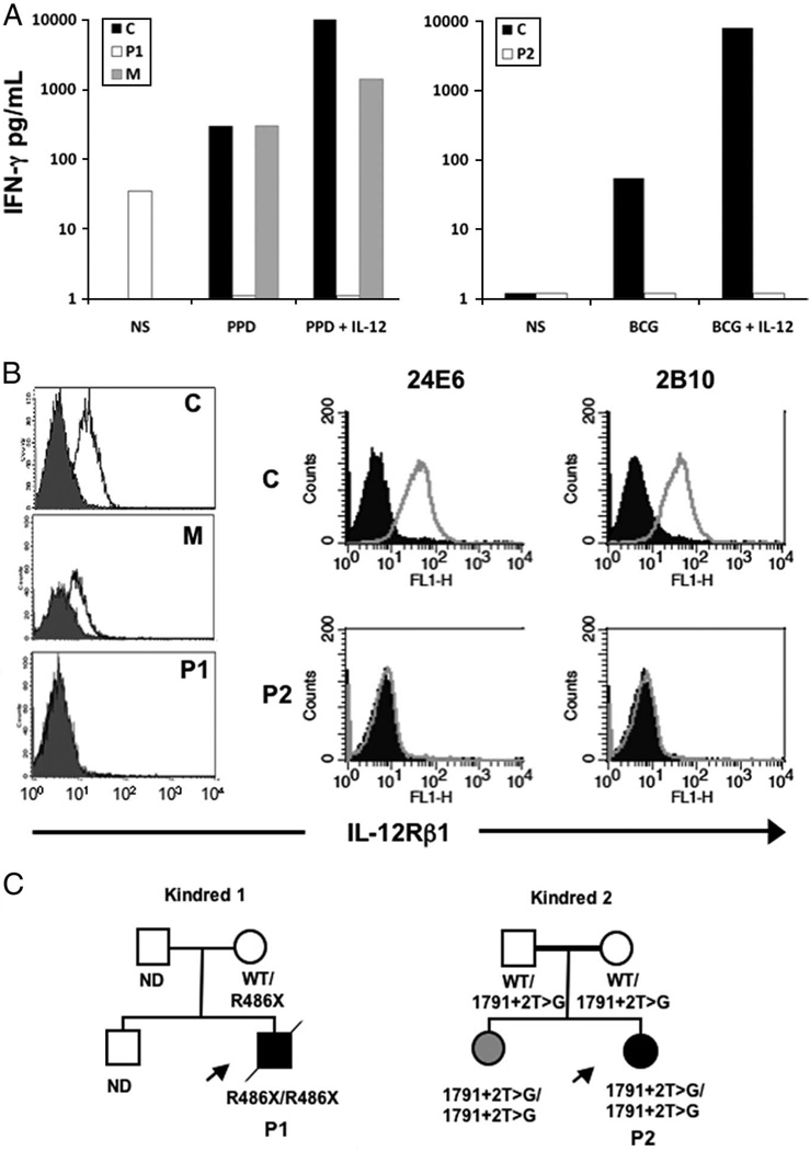
FIGURE 1.
A, IFN-γ produced by stimulated whole-blood samples was undetectable in the patients, and they did not respond to IL-12. NS indicates not stimulated; P1, patient 1; P2, patient 2; M, mother of patient 1; PPD, purified protein derivative from M tuberculosis; C, healthy control. B, IL12Rβ1 was not expressed on PHA-T blasts, as assessed with 1 (patient 1, left) and 2 (patient 2, right) monoclonal antibodies. FL1-H indicates ●●●●●●; open histograms, IL12Rβ1; closed histograms, isotype controls. C, Family trees of the studied patients showing the mutations found. ND indicates not done; WT, wild type; arrows, patients; diagonal line, the person concerned is dead.

