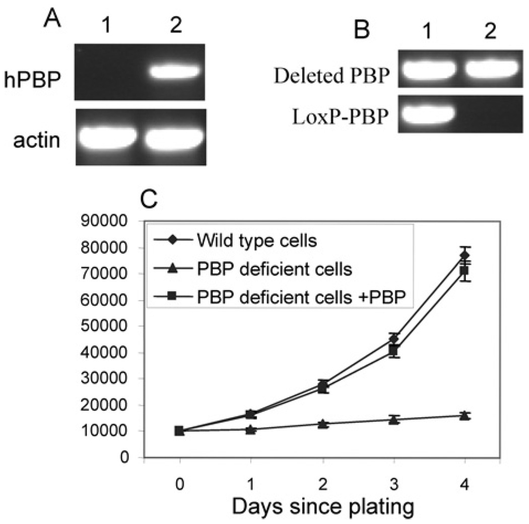
Figure 3. Restoration of PBP reversed the growth inhibition.
(A) Expression of PBP in PBP conditional knockout cells by transfection. PBPLoxP/LoxP Notch4-immortalized cells expressing Cre-ERtam were transfected with a control vector (lane 1) or PBP expression vector (lane 2). RT–PCR was performed to show the expression of human PBP (hPBP). (B) The PBP gene was mutated in cells transfected with the PBP expression vector but not in cells transfected with the control vector. PCR was performed with primers specific for the LoxP-PBP transgene and deleted PBP gene (Lane 1, +control; lane 2, +pcDNA3.1-PBP). (C) Vigorous growth of PBP knockout cells expressing exogenous PBP. Cells transfected with PBP expression vector (PBP deficient cells + PBP), cells transfected with the control vector (PBP deficient cells) or wild-type Notch4-immortalized cells were treated with tamoxifen for 2 days and then plated and counted at the indicated time points. Unlike PBP-deficient cells, the proliferation of PBP-deficient cells expressing exogenous PBP was not inhibited (P < 0.001).

