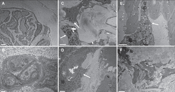
Figure 2).
Electron micrographs of varicose veins. Unmyelinated C fibres are identified between the media and adventitia (A and B) in patients 5 and 9, respectively. Mast cells (arrows) are present in the media of patient 2 (C and D); one of these mast cells is degranulated (D). A histiocytic macrophage overloaded with products of degradation (arrow) is in the vicinity of smooth muscle fibres in patient 6 (E). Smooth muscle fibres of the media from patient 2 appear disintegrated into pieces and scattered in an abundant collagen accumulation (F). Scale bar = 2 μm in A, C, E and F; scale bar = 200 nm in B; scale bar = 1 μm in D

