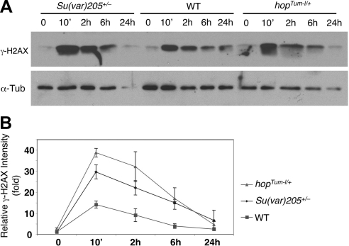
Figure 4.
Time course of radiation-induced DNA damage. A) Embryos of indicated genotypes were irradiated with 4000 rad of γ-rays or not irradiated. Total protein was extracted at different time points after irradiation (indicated), subjected to SDS-PAGE, and blotted with anti-γ-H2AX (14) and anti-α-tubulin (loading control). Results shown are representative of 3 independent experiments. B) Quantification of γ-H2AX vs. tubulin (loading control) for the indicated genotypes at different time points after irradiation as shown in panel A from 3 independent experiments. Note that Su(var)205+/− (Su(var)2055/+) and hopTum-l/+ animals exhibit greater levels of DNA damage than wild-type control animals (ry506)/w1118) at 10 min after irradiation. Error bars = sd.

