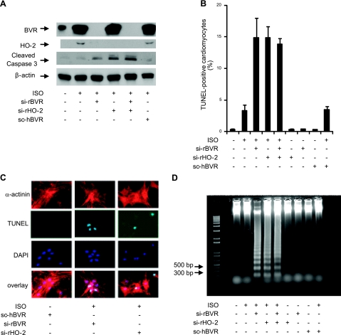
Figure 5.
Inhibition of BVR and HO-2 protein expression during ISO stimulation increases cardiomyocyte apoptosis. A) Cardiomyocytes were treated with siRNA against rat BVR, HO-2, or both, after which ISO was added at a final concentration of 10 μM, and the cells were incubated for 24 h. A randomized form of sihBVR (scBVR) was added to some cells as a control. Expression of BVR, HO-2, and cleaved caspase-3 was determined by Western blot. β-Actin served as a loading control. B) Quantitative analysis of the number of apoptotic cells detected, in cultures treated with siBVR, siHO-2, or both; scBVR served as control. Number of TUNEL-positive cells that also stained with EA-53 anti-α-actinin antibody was measured as a fraction of all cardiomyocytes. C) Detection of apoptosis in cardiomyocytes by microscopy. Cardiomyocytes were characterized by their staining with antibody against α-actinin. Apoptosis was detected by TUNEL staining; nuclei were visualized with DAPI. D) Total genomic DNA was isolated from cardiomyocytes that had been treated with ISO and siBVR, siHO-2, or both. Fragmentation of nuclear DNA is detectable in ISO-treated cardiomyocytes that were also treated with siBVR, siHO-2, or both. The 1-kb-plus ladder was used as DNA marker.

