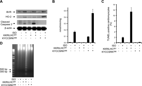
Figure 6.
Peptide-mediated inhibition of BVR activation in perfused heart increases cardiomyocyte apoptosis, whereas BVR activation is protective. A) Three groups of 4 rats were injected subcutaneously with ISO (0.01 mg/kg), twice, with a 6-h interval between injections prior to perfusion of the isolated hearts. As indicated, isolated hearts were perfused with the peptides (25 μM) KKRILHC281 or KYCCSRK296 in the presence or absence of 0.1 μM ISO, for 3 h. Levels of BVR, HO-2, and cleaved caspase-3 were determined by Western blot, using β-actin as a control for equal loading. B) BVR activity was measured at pH 6.7, using NADH as cofactor. Rate of conversion of biliverdin to bilirubin was determined from the increase in absorbance at 450 nm at 25°C. Specific activity is expressed as nanomoles of bilirubin per minute per milligram of protein. C) Detection of apoptosis in perfused heart cardiomyocytes. Tissue from the perfused hearts was analyzed using the TUNEL assay, as described in Fig. 5B. D) Genomic DNA was isolated from perfused heart, and resolved by agarose gel electrophoresis; fragmentation is detectable only in hearts perfused with ISO and the inhibitory peptide, KKRILHC281. The 1-kb-plus ladder DNA was used as a marker.

