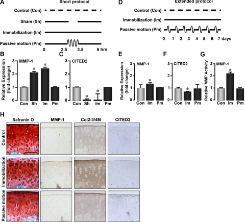
Figure 1.
Passive motion loading prevents cartilage degradation. Schematics: representations of the short protocol (A), where the rat hind limb is immobilized for 6 h interrupted by 1 h of passive motion or release from immobilization under anesthesia, or the extended protocol (D), where the rat limb is immobilized for 7 d with or without a 1-h/d passive motion protocol. Graphs: qPCR showing fold-change in mRNA levels of MMP-1 (B, E) or CITED2 (C, F), and MMP-1 (enzyme) activity (G) after immobilization (Im), passive motion loading (Pm), sham treatment (Sh), or no treatment (Con). Images: Safranin O staining and immunohistochemical localization of MMP-1, type II collagen denaturation (Col2–3/4M antibody), and CITED2 in articular cartilage from rats undergoing the extended protocol (H). For statistical analysis, 1-way ANOVA and Tukey's post hoc test were performed; n = 5 rats/group. Scale bar = 100 μm. *P < 0.05 vs. control.

