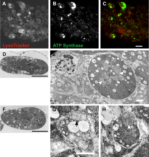
Fig. 9.
Mitochondrial clusters colocalize with LysoTracker staining and can be seen in membrane-bound organelles by TEM. (A-C) A degenerating egg chamber stained with (A) LysoTracker and (B) anti-ATP synthase. (C) Overlay shows mitochondrial clusters (green) in LysoTracker-positive regions (red). Scale bar: 10 μm. (D,F) Degenerating wild-type stage 8 egg chambers. Sections (1 μm) stained with 1% Toluidine Blue showing overall morphology of egg chambers. Scale bars: 50 μm. (E) Electron micrograph of a region of the degenerating egg chamber in D. (G,H) Electron micrographs of the degenerating egg chamber in F. Arrow in G indicates a degenerating mitochondrion. Arrowheads in E and H indicate mitochondria. Scale bars: 1 μm in E,G.

