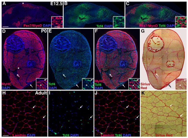
Fig. 1.
Tcf4 is highly expressed in muscle connective tissue fibroblasts. In the embryonic (A-C), neonatal (D-G) and adult (H-K) limb, Tcf4+ fibroblasts are associated with Pax7/MyoD+ myoblasts (A-C), MyHC+ myofibers (D-F) and laminin-ensheathed myofibers (H-J). By P0 and in the adult mouse, Tcf4+ fibroblasts (arrows) lie within the Sirius Red+ connective tissue (G,K) outside the MyHC+ myofibers (D-F) and laminin+ muscle basal lamina (H-J). In the neonate, some myonuclei (arrowheads) express low levels of Tcf4. Asterisks in A and D indicate enlarged inset regions in A-G. Scale bars: in A, 200 μm in A-G; in A inset and H, 50 μm in A-G insets and H-K.

