Fig. 3.
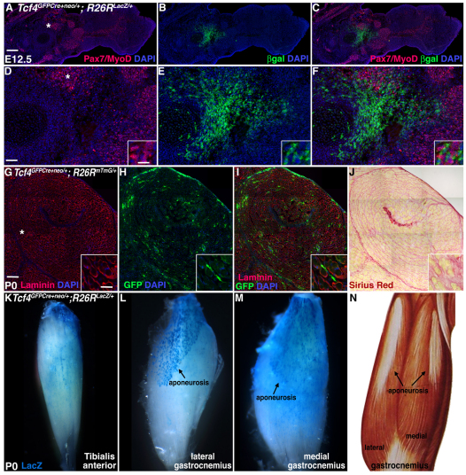
Tcf4GFPCre+neo genetically labels muscle connective tissue fibroblasts in vivo. (A-J) In the embryonic limb of Tcf4GFPCre+neo/+;R26RlacZ/+ mice (A-F) and neonatal limb of Tcf4GFPCre+neo/+;R26RmTmG/+ mice (G-J) β-gal+ (A-F) or membrane bound GFP+ (G-I) Tcf4-derived cells lie associated with, but interstitial to, Pax7/MyoD+ myoblasts (A-F) or laminin-ensheathed myogenic cells (G-I) and in Sirius Red+ regions (J). (K-M) In whole-mount preparations, β-gal+ cells (blue) are present throughout the tibialis anterior and gastrocnemius muscles and concentrated in the gastrocnemius aponeurosis of Tcf4GFPCre+neo/+;R26RlacZ/+ mice. (N) Drawing of the gastrocnemius muscle. Asterisks in A, D and G indicate enlarged regions in D-F, insets in D-F and insets in G-J, respectively. Scale bars: in A, 50 μm for A-C; in D, 12.5 μm for D-F; in D inset, 3.125 μm for D-F insets; in G, 100 μm for G-J; in G inset, 25 μm for G-J insets.

