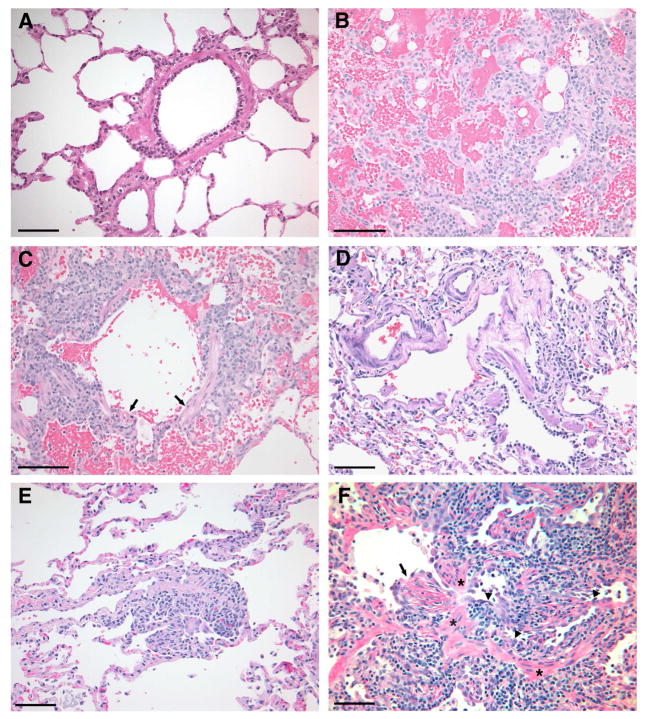
Figure 2. Necropsy histology and rejection classification of the lung grafts at the end of study (see Table 2 for a description of the grading system).
A) Autologous control (dog G135, group 1): Normal lung histology with no evidence of graft rejection (Grade A0, B0).
B) Rejection control (dog G533, group 3): The vessel in the lower right corner displays a perivascular lymphoid infiltrate which extends into the alveolar septal interstitium. There is marked alveolar hemorrhage. The alveolar septal devitalization in the upper left corner borders an area of parenchymal necrosis. (Grade A4).
C) Rejection control (dog G533, group3): In the same lung illustrated in (B), there is peribronchial lymphocytic infiltration and focal necrosis of bronchiolar epithelium (arrows). The lymphoid infiltrate extends into the alveolar interstitium. Alveolar hemorrhage is present. (Grade B4).
D) Chimeric recipient (dog G190, group 2): Normal lung histology with no evidence of graft rejection at end of study (Grade A0, B0; 27 months).
E) Chimeric recipient (dog G005, group 2): This perivascular infiltration by small numbers of lymphocytes is consistent with mild graft rejection. The airways (not shown) were uninvolved. (Grade A1, B0; 29 months).
F) Chimeric recipient (dog E875, group 2): Histological changes are observed consistent with active acute and chronic graft rejection (Grade A2, B1, Ca; 27 months). The bronchiole which extends horizontally across the photomicrograph is narrowed by lymphocytic infiltration of the mucosa (arrowheads) and proliferation of granulation tissue (arrow). Bands of smooth muscle (asterisks) in the bronchiolar wall serve as anatomic landmarks. The alterations are characteristic of early obliterative bronchiolitis, grade Ca (active) in the International Society for Heart and Lung Transplantation grading system, This is the histological expression of active chronic lung rejection as well as chronic pulmonary graft-versus-host disease. Bars represent 100 microns in all panels. Images were processed using Photoshop 7.0 software (Adobe Systems, San Jose, CA).

