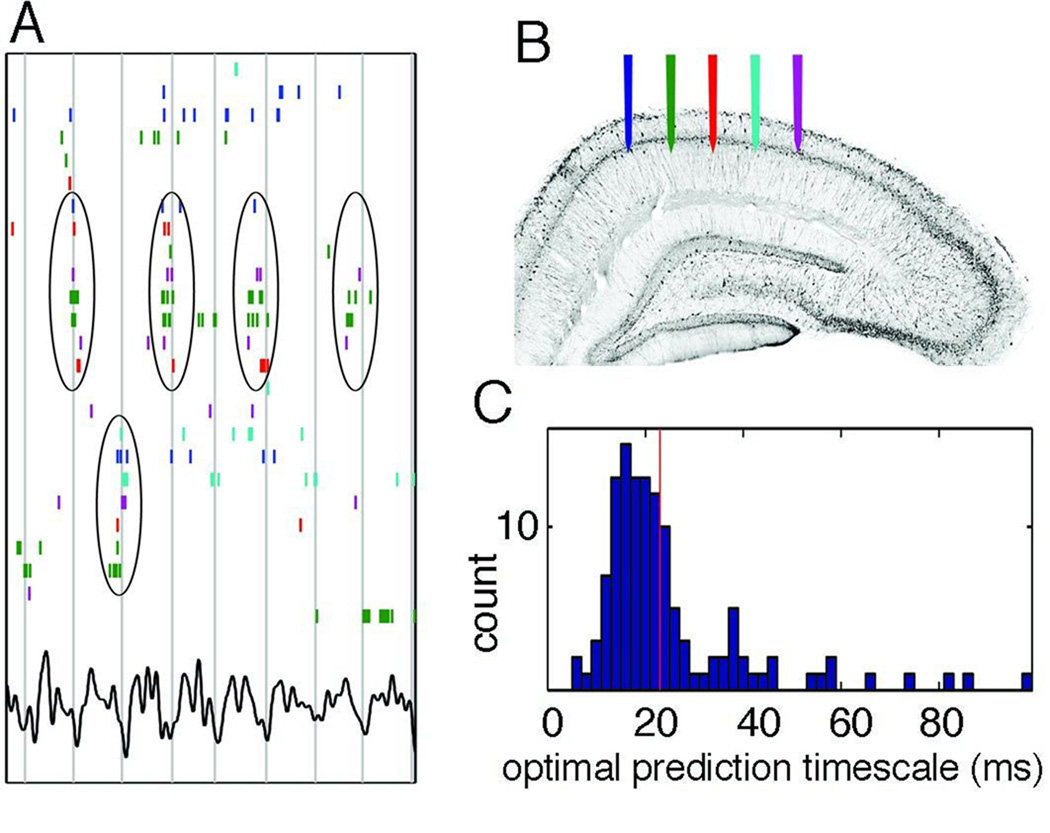
Figure 2.
Cell assembly: the fundamental unit of neural syntax. (A) Raster plot of a subset of hippocampal pyramidal cells that were active during a 1-s period of spatial exploration on an open field out of a larger set of simultaneously recorded neurons, ordered by stochastic search over all possible orderings to highlight the temporal relationship between anatomically distributed neurons. Color-coded ticks (spikes) refer to recording locations shown in (B). Vertical lines indicate troughs of theta waves (bottom trace). ‘Cell assembly’ organization is visible, with repeatedly synchronous firing of some subpopulations (circled). Note that assemblies can alternate (top and bottom sets) rapidly across theta cycles. (C) Spike timing is predictable from peer activity. Distribution of time scales at which peer activity optimally improved spike time prediction of a given cell, shown for all cells. The median optimal timescale is 23 ms (red line). Modified after Harris et al. (2003).

