
Figure 6.
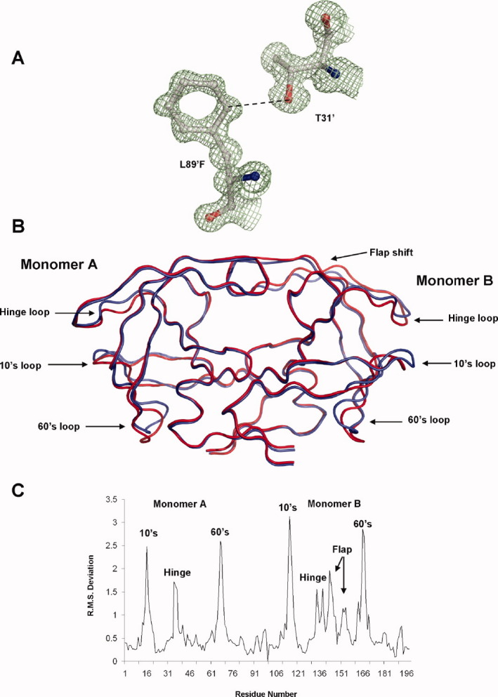
A: 2Fo − Fc electron density map contoured at 1.5σ for L89′F polymorphic substitution in monomer B of PR1N/DRV complex. The shortened CH…O interaction with Thr31′ is indicated by the broken line. B: Superposition of PR1N dimer in red against PR1M dimer in blue. The core of the PR1N structure including the active site shows excellent agreement with the PR1M structure. The two structures deviate in the 10s loop, 60s loop, and the flap hinge regions where the majority of polymorphic substitutions map. The flap of monomer A agrees well with that of PR1M structure, but the flap of monomer B deviates by ∼1 Å. C: The RMS deviations (Å) per residue when compared with those of PR1M are plotted for the Cα atoms of PR1N. The two monomers are numbered 1–99 and 101–199. The deviations corresponding to 10s, 60s, and hinge loops for both the monomers and the flap of monomer B are indicated. An interactive view is available in the electronic version of the article. PRO486 Figure 6