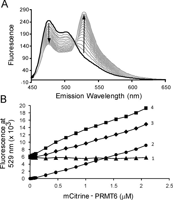
Figure 4.

mCerulean and mCitrine-PRMTs produce FRET. (A) A 1.0-μM solution of mCer-PRMT1 is added to a cuvette to a final volume of 1.5 mL. mCit PRMT1 is then titrated for sixteen 10-μL additions into the sample, covering a concentration range of 0–3.5 μM. After each addition, the solution is allowed to stir for 2 min prior to scanning for wavelength emission between 450 and 650 nm using a Varian benchtop fluorometer as described in the Materials and Methods section. (B) The emission at 529 nm is measured using a Biotek micro-plate reader as described in the Materials and Methods section. The background fluorescence from 0.5-μM mCer-PRMT6 alone (▴) remains constant, and the background fluorescence contributions from 0- to 2.08-μM mCit-PRMT6 alone (•) increases linearly with increasing protein. The combination of a fixed concentration of mCer-PRMT6 (0.5 μM) with varying concentrations of mCit-PRMT6 (▪) shows greater fluorescence intensity than the sum of both background signals (♦).
