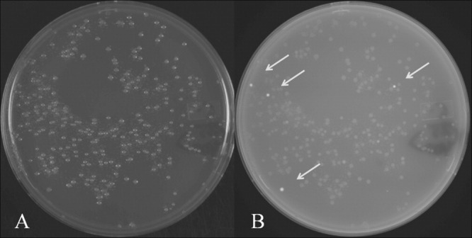
Figure 1.

A typical chimeric-GFP solubility screening experiment. (A) white light image, (B) transilluminating UV (365 nm) image of an agar plate used to select GFP-positive colonies in the solubility screen. Electrocompetent DH10B cells were transformed with a transposase fragment library, which was subcloned as a chimera into the GFP-containing vector pPROGFP. The right image contains four “bright” GFP-positive colonies (arrows) which are selected for expression testing, sequencing, and solubility studies. The sensitivity of the screen can be modulated by early or late detection of GFP-positive clones.
