
Figure 3.
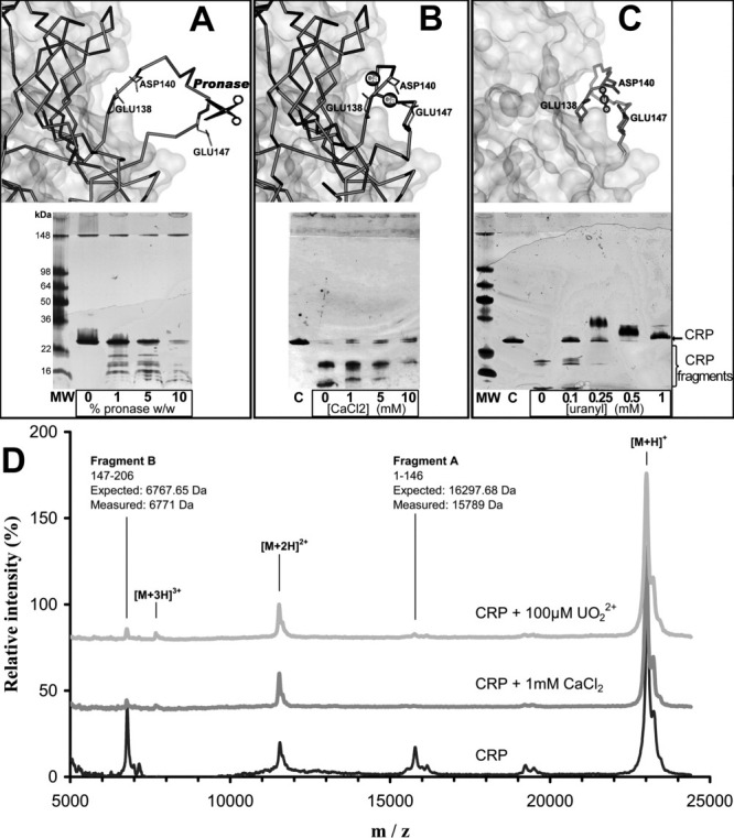
Conformational sensitivity of CRP to digestion with pronase. A: Top: without bound calcium atoms, the CRP138–148 loop is largely exposed to solvent (IGNH chain D). Bottom: electrophoresis gel of CRP (200 μg/mL) digestion at a fixed CaCl2 concentration (1 mM) and an increasing concentration of pronase, which promotes CRP (23 kDa) digestion. B: Top: in the presence of two bound calcium atoms, the CRP138–148 loop is less exposed to the solvent (1GNH chain C). Bottom: electrophoresis gel of CRP (200 μg/mL) digestion at a fixed concentration of pronase (5%W/W) and an increasing concentration of CaCl2. Calcium precludes CRP digestion by pronase41. C: Top: predicted binding site geometry of chelated  geometry on CRP chain C (IGNH). The side chains responsible for calcium chelation in CRP are also involved in binding. Bottom: protection against pronase digestion of CRP (200 μg/mL) with increasing concentrations of the uranyl ion. Electrophoresis results represent at least two independent silver staining experiments. D: MALDI-TOF mass spectra of CRP. CRP was incubated without metal (black), with 1 mM CaCl2 (dark grey) and 100 μM (light grey) prior to pronase digestion. Relative intensity was set at 100% for the [M+H]+ peak. [M+H]+, [M+2H]2+, and [M+3H]3+ are assigned to mono, doubly, and triply charged intact CRP peaks, fragments A and B are pronase digestion products. Calcium and spectra show very similar peak distribution, with near disappearance of digestion products in agreement with electrophoresis results, implying that the conformation of the metal-bound CRP loop is similar for both metals.