
Figure 3.
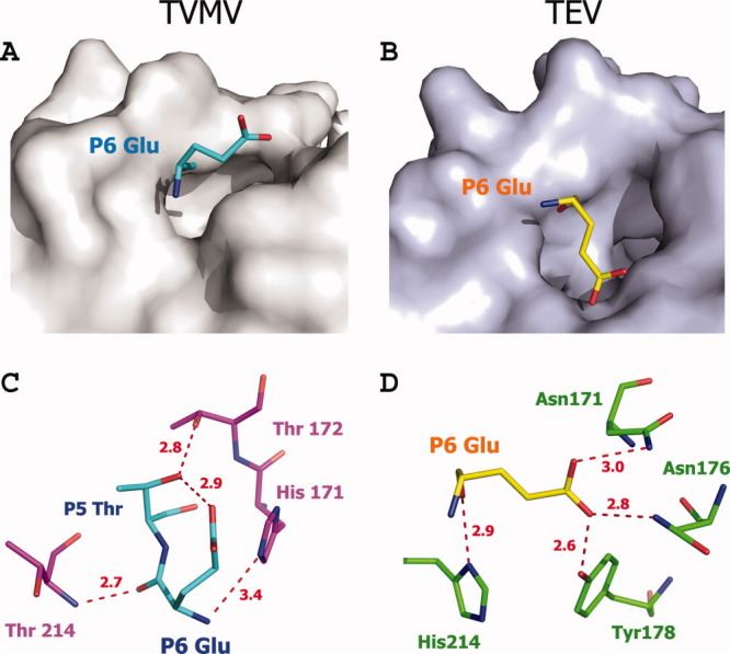
Comparison between the P6 positions of TVMV and TEV protease (PDB ID: 1LVB, Chain A) substrates. (A, B) P6 Glu binds to the surface of its corresponding protease, viewed at the same angle. (C, D) Hydrogen-bond interactions between P6 Glu and TVMV protease (C) and TEV protease (D). Residues are shown in ball-and-stick representation. The hydrogen bonds are shown as dashed lines colored red. Residues from TVMV and TEV protease are colored in purple and green, respectively. The P6 substrate residues for TVMV and TEV proteases are colored cyan and yellow, respectively.