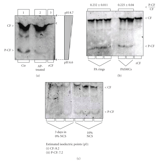
Figure 1.
Assay of the phosphocofilin (P-CF) content in canine PA rings and cultured PASMCs by IEF. Total protein from freshly dissected PA rings (panel (a)) and undifferentiated and differentiated cells (panel (b)); recombinant CF control (rCF) were resolved by IEF electrophoresis on gels containing 1% of each ampholyte pH 6/8 and 7/9. For CF dephosphorylation, protein extracts were incubated with calf intestinal alkaline phosphatase (AP) prior to IEF electrophoresis. Protein was transferred onto nitrocellulose membranes and membranes were probed with a cofilin antibody that recognizes both unphosphorylated (CF) and phosphocofilin (P-CF). Immunoreactive bands were quantified by densitometry, and treatment-dependent changes of the P-CF/CF ratio were presented relative to untreated controls (Ctr). Vertical strips of gels (0.5 cm wide) were cut into 1 cm long segments and soaked in deionized water before assay of pH (pH bar in panel A). Panel (c): CF expression is upregulated in proliferating PASMCs (cultured in 10% NCS), compared to differentiated cells (cultured for 4 days in 0% NCS).

