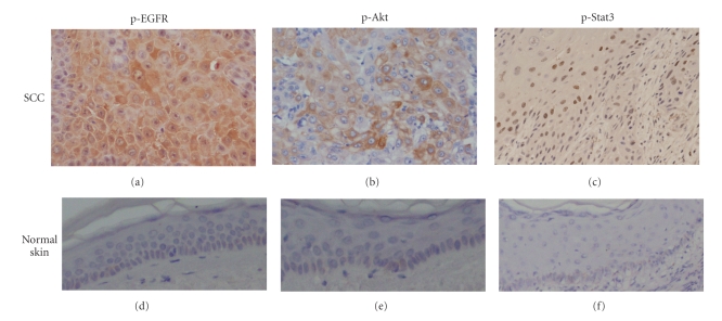
Figure 1.
Over-expression of p-EGFR, p-Akt, and p-Stat3 in human cutaneous SCC tissues. Paraffin-embedded serial sections of SCC from the same patient were examined immunohistochemically for p-EGFR, p-Akt, and p-Stat3 (tyr705) expression. Characteristic immunostaining of p-EGFR, p-Akt and p-Stat3; original magnification: ×200. P-EGFR (a) and p-Akt (b) protein were overexpressed mainly in the cytoplasm and partially in the nucleus. P-Stat3 (tyr705) protein was detected in the nuclei (c). None of p-EGFR p-Akt or p-Stat3 protein were seen in normal skin ((d), (e), (f)); original magnification: ×100.

