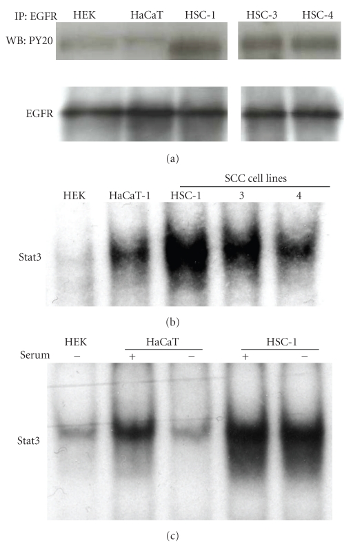
Figure 3.
Analysis of EGFR and Stat3 activation in human epidermal primary keratinocytes (HEK), HaCaT cells, and human SCC cells. Cells were incubated in serum-free medium for 24 hours, and then cell lysates were extracted from HEK, HaCaT cells, HSC-1, -3, or -4 cells. Total 100 μg of protein in each lysate were subjected to immunoprecipitation (IP) with the anti-EGFR antibody. The immunoprecipitates were separated by SDS-PAGE and subjected to immunoblot analysis with an anti-phosphotyrosine antibody, PY20. The same membrane was then reprobed with the anti-EGFR antibody. Data are representative of three independent experiments (a). Nuclear extracts from HEK, HaCaT cells, HSC-1, -3, or -4 cells under the regular condition were analyzed using EMSA with a 32P-labeled DNA probe to detect DNA binding activity of Stat3 (b). HSC-1 or HaCaT cells were seeded in medium with 10% fetal calf serum, and the cells were cultured for 24 hours, after which they were cultured in medium with or without 10% fetal calf serum for 24 hours and Stat3 activation was analyzed using EMSA (c).

