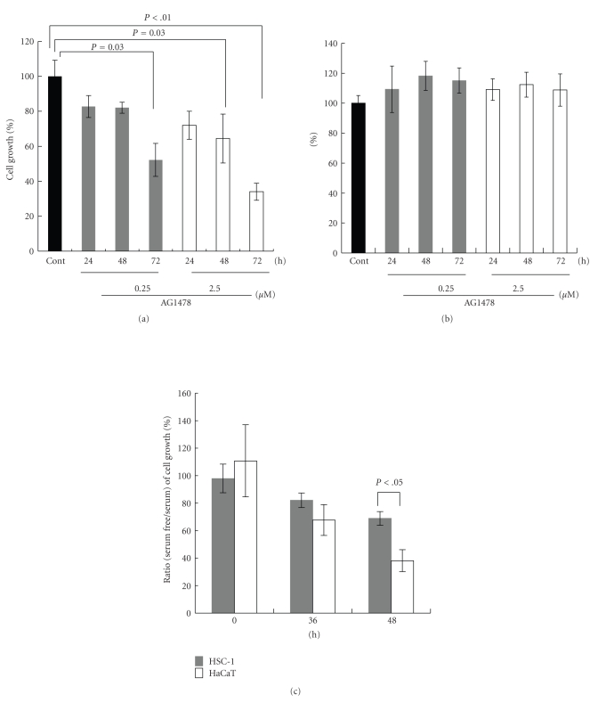
Figure 5.
Effect of AG1478 on proliferation of HSC-1 cells and HEKs. After HSC-1 cells or HEKs were incubated with AG1478 (0.25 or 2.5 μM) for indicated periods, cell proliferation was analyzed using the MTS assay ((a), (b)). Cell growth of HSC-1 cells (a) or HEKs (b) was expressed as a percentage of that in untreated cells at each indicated time point up to 72 hours. HSC-1 or HaCaT cells were seeded in medium with 10% fetal calf serum and were cultured for 24 hours. The medium was then changed to serum-free medium or medium with 10% fetal calf serum. The growth of HSC-1 and HaCaT cells was monitored using the MTS assay at 0 hour, 36 hours, and 48 hours after the change of medium and was expressed as the percent ratio (serum-free/serum) (c). Data are mean ± SD of at least three experiments. P < .05 when compared with each group.

