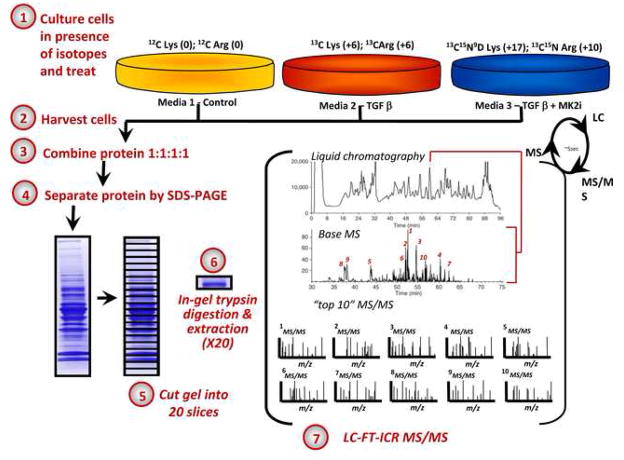
Figure 1.
Schematic of workflow used for a three-way SILAC-MS experiment. Three cell populations are isotopically labeled with normal and stable isotope-substituted arginine (R) and lysine (K) amino acids, creating three cell populations distinguishable by mass. Each population is stimulated as indicated yielding 3 samples which are combined in equal amounts (25 μg each), trypsin-digested, and analyzed using mass spectrometry.

