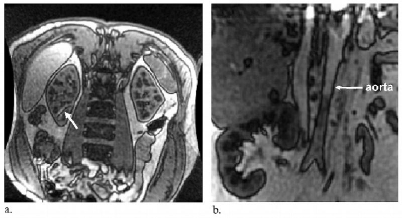
Figure 2.

(a) Worst case example of b-SSFP banding artifact inside the kidney for a healthy native kidney subject. The nulled banding area (white arrow) is excluded from mean cortical perfusion calculations. (b) Intersection of the imaging slice with major feeding vessels, such as the aorta in this example (white arrow), prevented a coronal acquisition in many of the transplant subjects. In these cases, a coronal acquisition would have caused the slice selective inversion to invert the inflowing blood spins which are presumed to be at equilibrium magnetization upon entrance to the kidney.
