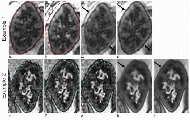
Figure 3.

Two different transplanted kidneys which demonstrated significant motion during the ASL exam. Example 1 displays translation due to respiratory motion (dotted outline in a-b). The average of the two images pre-registration demonstrated motion artifact at the boundary of the kidney body and blurring (arrow in c), while averaging post-registration mitigated these (arrow in d). Example 2 displays another transplanted kidney in three different positions (e-g). The average of all 64 FAIR images pre-registration demonstrated blurring (arrow in h) which the post-registration average reduced (arrow in i).
