Figure 4.
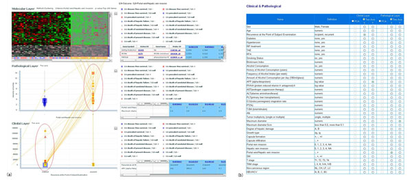
2 Dimensional 3 Layered map. Two dimensional three layered map, composed of molecular, pathological and clinical layer map, shows the relative position of the patient within each layer and interrelation between the position of different layer. As shown in this figure, the user can intuitively understand the relation among gene expression pattern (molecular state), pathological state and clinical state of the patient. The axes of the pathological or clinical layer are the first and the second principal component extracted from the principal component analysis applied to the user-specified group of pathological or clinical items, respectively. (a) In the molecular layer, overexpressed genes are chosen, corresponding to the criterion "Portal vein/Hepatic vein invasion +". We can find MCM6 gene, DNA replication licensing factor in the overexpressed genes. On the other hand, pathological layer shows that larger tumors are found (see x-axis of pathological layer) from among this group. Lines between pathological layer and clinical layer show that “incipient” cases (x-axis in clinical layer) usually have larger tumors. (b) Parameter settings, used to draw the map. This system can use the axis specified by the user. In clinical layer, x-axis is recurrence, y-axis is AFP (tumor markers for detection of hepatocellular carcinoma). In pathological layer, x-axis is "Portal vein/Hepatic vein invasion”, y-axis is “Maximum Diameter”.

