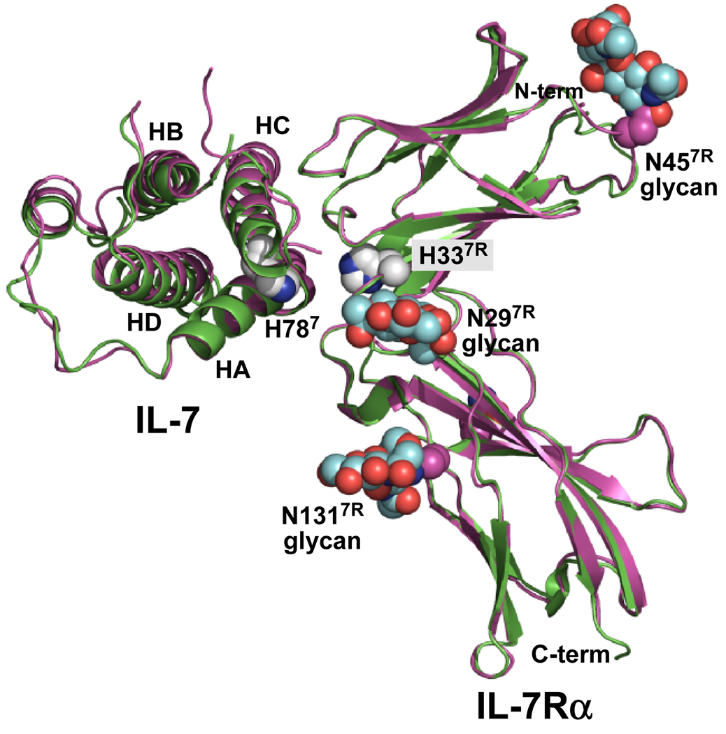
Figure 1.
Ribbon diagrams of the complex structures of IL-7 bound to nonglycosylated (green, 3di2.pdb) and glycosylated (magenta, 3di3.pdb) forms of the IL-7Rα (7). The nonglycosylated complex was superimposed onto the glycosylated complex. The 4 α-helical bundle of IL-7 is labeled helix A (HA), B (HB), C (HC), and D (HD) accordingly. The N-glycans attached to N297R, N457R, and N1317R are colored as cyan CPK groups for the carbon atoms. The only histidine residues observed in the IL-7/IL-7Rα interface involve H787 and H337R. The histidine side chains are drawn as white CPK groups for the carbon atoms. Oxygen and nitrogen atoms are colored red and blue, respectively. This picture was created and rendered using PyMOL (58).

