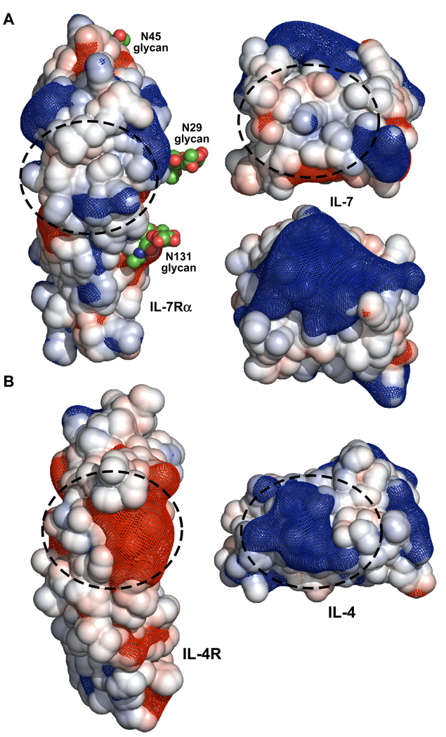
Figure 7.
Open book view of the electrostatic surfaces and potentials for the IL-7/IL-7Rα (A) and IL-4/IL-4Rα (B) complexes. The linear Poisson-Boltzmann equation was solved for both protein complexes using APBS (33) with 150 mM monovalent salt at 298 K. The solvent accessible surface areas for each protein are displayed and colored blue (+5 kT/e) and red (−5 kT/e). The electrostatic potential gradients for each protein are displayed as a blue (+2 kT/e) and red (−2 kT/e) mesh. The structures and electrostatic potentials were displayed using PyMOL (58). The binding interfaces for both complexes are highlighted with dashed circles. The second view of IL-7 below the view with the dashed circle is rotated 90° in the vertical direction to illustrate the large positive surface on the top of the cytokine.

