Figure 3.
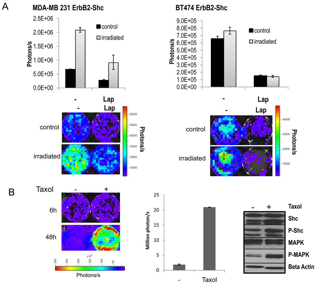
Monitoring erbB2/Her2 reporter activity in cancer cells exposed to cytotoxic treatments.
A). Her2-luc reporter activity in response to irradiation in MDA231-ErbB2-shc cells (left) and BT474-ErbB2-shc cells (right). Top panels: quantitative analysis of luciferase signals 72h after cells had been subjected to irradiation with 5Gy. Lower panels: representative bioluminescent images of irradiated (5Gy) and non-irradiated cells at 72h after treatment.
B). Induction of the ErbB2 pathway in response to treatment with taxol at a concentration of 0.5uM. Luciferase signals (left panel) and quantitative analysis (middle panel) of MDA-MB231 ErbB2-Shc cells 6h and 48h after incubation with taxol. Middle panel: Quantitative analysis. Right panel: Western blot analysis of downstream targets of the ErbB2 pathway. Phospho-specific and non-specific antibodies directed against Shc and MAPK were used to assess the phosphorylation status of Shc and MAPK after taxol incubation for 48h. Error bars represent standard error of the mean of three individual experiments.

