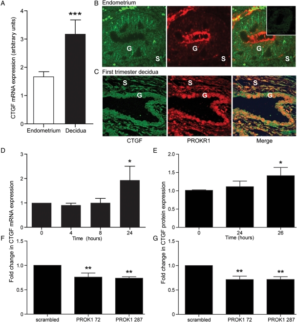
Figure 2.
CTGF expression and localization in human non-pregnant endometrium and first-trimester decidua and regulation by PROK1 in first-trimester decidua. (A) CTGF mRNA expression is elevated in first-trimester decidua (n = 42) compared with non-pregnant endometrium (n = 30). (B) In non-pregnant endometrium (n = 4; representative sections shown), double fluorescence immunohistochemistry demonstrated that CTGF (green panel) and PROKR1 (red panel) both localized to the glandular epithelium (G) and a subset of stromal cells (S). Negative control (−ve) is indicated in the figure insert. (C) CTGF and PROKR1 co-localized in the glandular epithelium (G) and a subset of stromal cells (S) in first-trimester decidua. Co-localization shown in merge panel (×20 magnification). Treatment of first-trimester decidua with 40 nM PROK1-increased CTGF mRNA (n = 10) (D) and protein expression (n = 7) (E), after 24 and 26 h of treatment respectively. Conversely, first-trimester decidua infected with lentivirus encoding miRNA constructs targeting PROK1 (72 or 287)-decreased CTGF mRNA (F) and protein expression (G) compared with control tissue infected with a scrambled miRNA sequence. ***P < 0.001, **P < 0.01, *P < 0.05 compared with non-pregnant endometrium (A), vehicle-treated tissue (D and E) or compared with scrambled siRNA PROK1 construct (F and G), as determined by Student's t-test (A) or one-way ANOVA (D–G).

