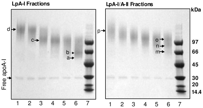
Figure 4. SDS-PAGE of cross-linked LpA-I and LpA-I/A-II sub-fractions.
Individual subfractions of LpA-I and LpA-I/A-II were cross-linked at 1:100 total protein to BS3 cross-linker ratio at 1 mg/ml protein concentration and were subjected to 4–15% SDS-PAGE. Band “b” on Lane 6 (Panel LpA-I) and band “m” on Lane 6 (Panel LpA-I/A-II) correspond to albumin contamination (~66kDa). The small amount of apoA-I that did not involve in intermolecular cross-linking is shown as “free apoA-I”. Labeling of fractions are as in Figures 2 and 3. Low Molecular Weight (LMW) markers are on Lane 7. Gels were stained with Coomassie Blue.

