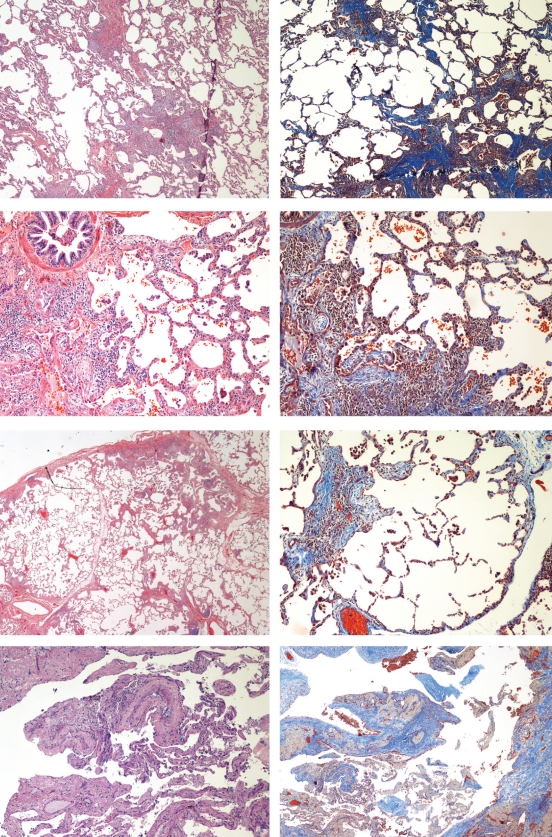
Figure 3.
Photomicrographs of hematoxylin and eosin- (left) and Masson’s trichromestained (right) sections. Proband (IV-10): top; III-2: second; III-4: third; and III-7: bottom. All show the usual interstitial pneumonitis pattern with patchy fibrosis, intervening normal lung parenchyma and fibroblastic foci

