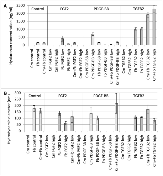
Figure 1. HA concentration and size in cell media.
Cultured cardiomyocytes, fibroblasts and co-cultured cells (80%/20%) was stimulated with growth factors (FGF2, fibroblast growth factor-2, PDGF-BB, platelet-derived growth factor-BB and TGFB2, transforming growth factor-β2). “Low” indicates a growth factor concentration in the media of 50 ng/mL for PDGF-BB and 5 ng/mL for FGF2 and TGFB2. “High” indicates a growth factor concentration in the media of 100 ng/mL for PDGF-BB and 10 ng/mL for FGF2 and TGFB2. HA concentration was measured with an enzyme-linked binding protein assay. HA size was estimated with DLS.

