
Fig. 1.
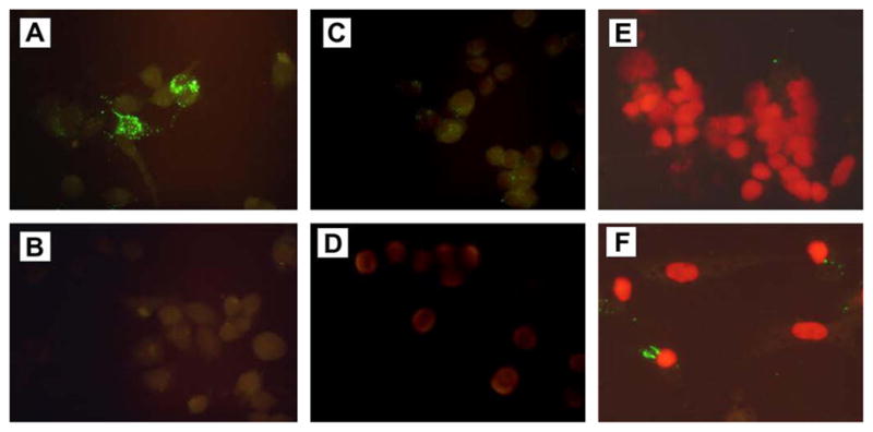
Uptake of S41 phage into mouse neuroblastoma N2A cells and rat cerebellar granule neurons (CGNs) detected by immunofluorescence. Panels show immunofluorescence (IFA) detection of internalized phage in N2A cells (A–D) and CGNs (E,F). Cells were incubated with S41 phage (A, B and F), control phage (C) or remained untreated (D,E). IFA was performed as described in “Materials and methods”. Cells were stained with FITC-labeled secondary antibody in the presence (A, C, D, E, and F) or absence (B) of mouse anti-M13 mAb. All cells were stained with FITC-labeled anti-mouse IgG and counterstained with propidium iodide (PI) to show the nuclei prior to microscopic examination.