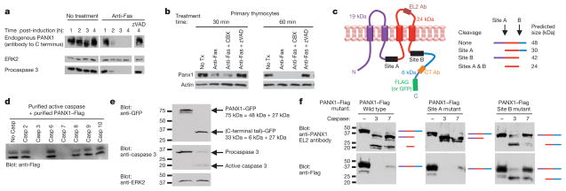
Figure 4. Pannexin 1 is a target of effector caspase cleavage during apoptosis.
a, b, Fas-mediated apoptosis results in loss of PANX1 detection with antibody targeted to PANX1 C terminus in Jurkat cells (a) and primary murine thymocytes (b). c, Schematic of PANX1–Flag protein indicating the predicted caspase cleavage sites (sites A and B) and the epitopes recognized by the EL2 (extracellular loop 2) and C-terminal antibodies. The predicted products of caspase cleavage at sites A and B are also shown. d, In vitro cleavage of immunoprecipitated PANX1–Flag incubated with the indicated purified active caspases. Loss of immunoreactivity to Flag assessed. Representative of two independent experiments. e, Jurkat cells stably expressing a C-terminally GFP-tagged PANX1 were induced to undergo apoptosis and assessed for the cleaved C-terminal fragment (which runs at ~33 kDa). Representative of two independent experiments. f, In vitro cleavage of indicated PANX1–Flag proteins (wild type and caspase cleavage site mutants), incubated with the indicated caspases.

