Figure 6.
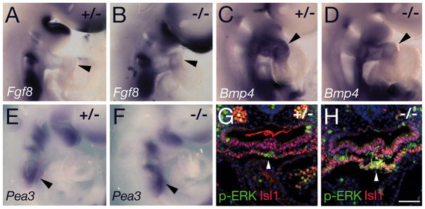
Analysis of signaling pathways in Tbx3 mutant embryos at E9.5. (A, B) In situ hybridization showing elevated levels of Fgf8 transcript accumulation in the OFT of Tbx3−/− (B) relative to Tbx3+/− (A) embryos (arrowheads). (C, D) Bmp4 transcripts are reduced in the OFT of Tbx3−/− (D) relative to Tbx3+/− (C) hearts (arrowheads). (E, F) Pea3 transcripts are slightly upregulated in the caudal pharynx of Tbx3−/− embryos (F, arrowhead). (G, H) Fluorescent immunohistochemistry showing elevated levels of nuclear phospho-ERK (green) in Isl1-positive ventral pharyngeal mesoderm (red) of Tbx3−/− (H, arrowhead) compared to Tbx3+/− embryos (G). Scale bar (G, H): 100μm.

