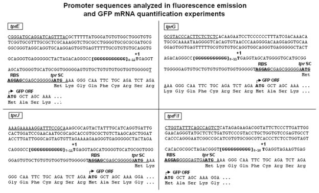
Fig. 2.
tprE, G, J and F/I promoter sequences cloned into the pGlow-TOPO vector and used for the GFP reporter assay. Regulatory elements are in bold. Underlining indicates the primers used to amplify the promoter regions. Numbers to the right of the homopolymeric repeats represent the number of G residues tested for each tpr promoter. As explained in Fig. 1, the tprE TSS (+1) is hypothetical and based on analogy with the other Subfamily II tprs. +1: transcriptional start site (experimentally determined); RBS: ribosomal binding site (putative); tpr SC: tpr gene start codon; GFP ORF: green fluorescent protein ORF. Amino acids (Lys–Gly–Gln–Phe–Cys–Arg–Ser–Arg) between the tpr putative SC and the GFP ORF start are vector-encoded.

