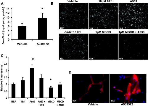
Fig. 3.
Inhibition of SCD-1 activity increases FC levels in MDA-MB-231 cells. In light of the fact that SCD-1 inhibition prevented cholesterol esterification with MUFA, we measured total cellular FC levels. A: cells were grown for 18 h in serum-free medium with or without 1 μM A939572, and FC levels were assessed by enzymatic assay. Inhibitor treatment caused a significant increase to total FC levels in culture. B: alternatively, cells were grown as in A and supplemented with indicated fatty acids, fixed, and stained with the FC probe filipin and visualized using fluorescence microscopy. C: inhibitor treatement increased filipin staining intensity and was reversed by cotreatment with 10 μm oleate or methyl-β-cyclodextrin (MβCD) treatment. Average fluorescence intensity was quantified from 10 randomly chosen fields from the images in B. D: excessive FC can alter membrane composition, and we saw that staining with the lipid raft probe cholera toxin B (red) and Hoescht (blue) increased with A939572 treatment. At ×10, scale bar represents 500 μm; at ×60, scale bar represents 50 μm.

