
Fig. 2.
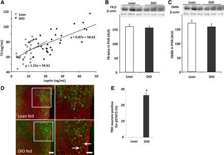
The change in the set point of the hypothalamic-pituitary-thyroid (HPT) axis of DIO rats correlates with plasma leptin and with an increase of pSTAT3 signaling within TRH neurons. A: correlation between serum leptin and serum tiiodothyronine (T3) in lean and DIO rats (r = 0.46 and 0.66 for lean and DIO rats, respectively). Each point represents a measurement from a single animal. B and C: thyroid hormone receptor-β2 (TRβ2) and leptin receptor (ObRb) protein levels were similar in both groups of rats. To perform this, hypothalamic paraventricular nuclei (PVN) were removed from fed lean and DIO rats; proteins were extracted in RIPA lysis buffer, and TRβ2 (60 kDa), ObRb (120 kDa), and β-actin (43 kDa) levels were assayed by Western blot. In addition, brain sections of fed rats were subjected to double immunohistochemistry using anti-pSTAT3 (brown staining) and anti-pro-TRH (red fluorescent staining) antiserum. D: low-magnification images for each individual staining for each group and high-magnification merged images. Arrows point to dual-labeled cells. Scale bars, 50 (low magnification) and 20 μm (high magnification). E: bar graph of the quantitative analysis of the percentage of TRH neurons positive for pSTAT3 in the PVN of fed lean and DIO rats (n = 3 and 5/group). Values are means ± SE. *P < 0.05 vs. lean group.