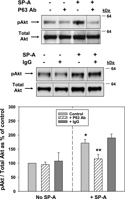
Fig. 10.
Activation of Akt by SP-A is inhibited by Ab to P63. Type II cells were serum-starved (4 h) and either were not (Control) or were preincubated with Ab against P63 (25 μg protein/ml, top set of gels) or nonimmune IgG (25 μg protein/ml, bottom set of gels) for 15 min before the addition of SP-A (1 μg/ml, 20 min). The cells were lysed and analyzed by Western blot as in Fig. 9. Samples from the same gel were cut as indicated and rearranged for clarity. Graph shows quantitation of gels in AU expressed as a percentage of control, no additions (mean ± SE, n = 3–8). *Significant difference from No SP-A Control; **significant difference from + SP-A control, P < 0.05.

