Fig. 8.
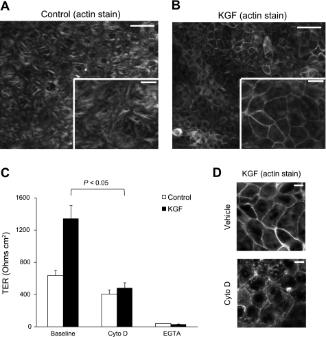
Effect of KGF administration on the actin cytoskeleton. Freshly isolated AT2 cells were plated on permeable supports in control media or media supplemented with KGF, and then cultured for 5 days, fixed, stained for F-actin with Alexa 568 phalloidin, and imaged under epifluorescence (A and B) or confocal microscopy (D). A and B: KGF-treated cells (B) demonstrated significant reorganization of the actin cytoskeleton with a marked increase in the apical perijunctional F-actin ring compared with control cells (A). Inset is higher magnification image (n = at least 4 biological replicates). Scale bars, 50 μm on ×20 image and 20 μm on inset ×40 image. C: actin depolymerization with cytochalasin D treatment decreased TER to control levels (P < 0.05) without a significant decrease in control TER (n = 4 biological replicates consisting of 4–6 technical replicates). Treatment of control and KGF-treated AT2 cells with calcium-free media resulted in near complete loss of TER. Data are expressed as means ± SE. D: the perijunctional F-actin ring in KGF-treated alveolar epithelial cells (top) was disrupted by treatment with cytochalasin D (bottom) (n = at least 4 biological replicates). Scale bars, 10 μm.

