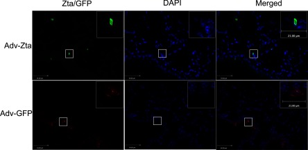
Fig. 1.
Immunofluorescent detection of Zta in the lungs of exposed mice. Histological sections from formalin-fixed, paraffin-embedded lung tissue of mice at day 7 postexposure were incubated with an antibody specific for Zta (top, left) or GFP (bottom, left). Visualization of antibody binding (green for Zta and red for GFP) was with a fluorescently tagged second antibody. The 2 right panels show blue nuclear DAPI staining of the same area. The boxed areas in the low-magnification images (×400) are shown magnified in the top, right corner inset (×630). The bar indicates 34 and 21 μm in the low- and high-power images, respectively.

