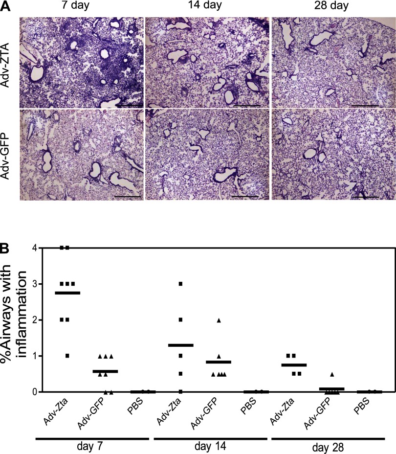
Fig. 4.
Resolution of lung inflammation in Adv-Zta-treated mice. A: mice were treated with 1 × 10⁸ pfu of the indicated adenovirus and killed at various times postexposure. The right lung of each mouse was fixed by intratracheal perfusion of 10% neutral buffered formalin. Hematoxylin and eosin (H&E)-stained lung sections from paraffin-embedded lung tissue of Adv-Zta (top) or Adv-GFP (bottom)-treated mice at 7 (left), 14 (middle), and 28 (right) days postexposure are shown. Scale bar represents 0.5 mm. B: histopathological assessment of lung inflammation at various times postexposure to adenovirus. H&E-stained lung sections shown in A from each mouse in the various treatment groups processed at the indicated times postexposure were evaluated for inflammation with the observer unaware of the sample identity. Scores for Adv-Zta (squares)-, Adv-GFP (triangles)-, and PBS (circles)-treated mice were assigned according to the percentage of airways associated with an influx of inflammatory cells (0 = 0–5%; 1 = 5–10%; 2 = 10–20%; 3 = 20–40%; 4 >40%).

